Introduction
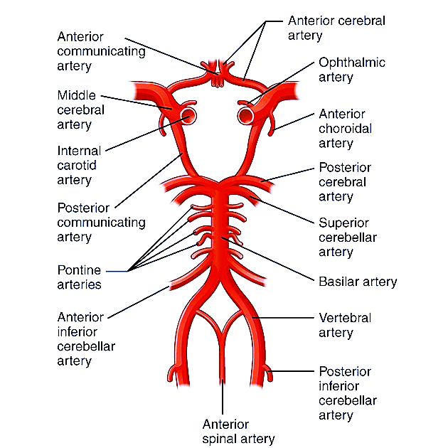
- The Circle of Willis is a arterial anastomotic ring located at the base of the brain, which connects the anterior and posterior cerebral circulations.
- Plays a critical role in maintaining collateral blood flow in cases of arterial occlusion.
- Location: Lies in the interpeduncular cistern of the subarachnoid space, surrounding the optic chiasm and pituitary stalk, resting on the ventral surface of the brain.
Components of the Circle of Willis
Anterior Circulation (supplied by Internal Carotid Arteries):
- Anterior cerebral arteries (ACA) – A1 segment (paired)
- Extends from ICA bifurcation to anterior communicating artery.
- Anterior communicating artery (ACOM) (single)
- Connects right and left ACA A1 segments.
- Terminal part of Internal Carotid Artery (ICA) (paired)
- Bifurcates into ACA and MCA.
- Posterior communicating arteries (PCOM) (paired)
- Connect ICA to posterior cerebral arteries.
Posterior Circulation (supplied by Vertebrobasilar System):
- Posterior cerebral arteries (PCA) – P1 segment (paired)
- Extends from basilar artery bifurcation to PCOM.
- Basilar artery tip (single)
- Formed by union of vertebral arteries.
Anatomical Arrangement
- Anterior half: Two ACA A1 segments connected by ACOM.
- Lateral connections: ICA terminal segment gives ACA & MCA; PCOM links ICA to PCA.
- Posterior half: Basilar artery bifurcates into PCAs, which join PCOMs to complete the ring.
- Shape: Hexagonal polygonal arterial network.
Arterial Territories Supplied
- ACA: Medial aspects of frontal and parietal lobes, anterior corpus callosum.
- MCA: Lateral hemispheric surface, basal ganglia (via lenticulostriate arteries).
- PCA: Occipital lobes, inferior temporal lobes, thalamus, and midbrain.
Common Variants (seen in ~50% of individuals)
- Hypoplastic segments – Most often PCOM or A1.
- Fetal-type PCA – PCA territory supplied predominantly by ICA via large PCOM; P1 is hypoplastic.
- Azygos ACA – Single midline ACA instead of paired A2 segments.
- Duplicated ACOM – Two parallel ACOMs instead of one.
- Persistent embryonic arteries – Persistent trigeminal, hypoglossal, or otic arteries connecting carotid and basilar systems.
- Unilateral absence of PCOM – May predispose to certain ischemic patterns.
Clinical Significance
- Collateral circulation: Maintains perfusion in case of proximal vessel stenosis or occlusion.
- Aneurysm formation: Most common site for berry aneurysms – ACOM > PCOM junction > MCA bifurcation.
- Ischemic stroke patterns: Distribution of infarcts can be predicted based on the vessel involved.
- Vasospasm: Common after SAH in the vessels of the circle.
Imaging in Radiology
- CT Angiography (CTA): First-line for assessing vessel anatomy and aneurysms.
- MR Angiography (MRA): Non-invasive; TOF-MRA useful for flow analysis.
- Digital Subtraction Angiography (DSA): Gold standard for aneurysm detection, vessel measurement, and pre-surgical planning.
- 3D Reconstructions: Aid in surgical and endovascular planning.
Diagram
Summary / Key Points
- Circle of Willis is a hexagonal arterial network at the brain base connecting anterior and posterior circulations.
- Made up of ACA (A1), ACOM, ICA, PCOM, PCA (P1), and Basilar artery tip.
- Highly variable – variants in ~50% population.
- Vital for maintaining cerebral perfusion in vascular compromise.
- Radiology: Best assessed with CTA, MRA, or DSA.