🔎 Introduction
Hydrocephalus is abnormal accumulation of cerebrospinal fluid (CSF) within the ventricular system, leading to increased ventricular size and sometimes raised intracranial pressure. Imaging plays a crucial role in diagnosis, classification (obstructive vs communicating), and surgical planning.
💻 CT in Hydrocephalus
Advantages
- First-line modality in emergency.
- Quick, widely available, excellent for ventricle size and hemorrhage detection.
CT Findings
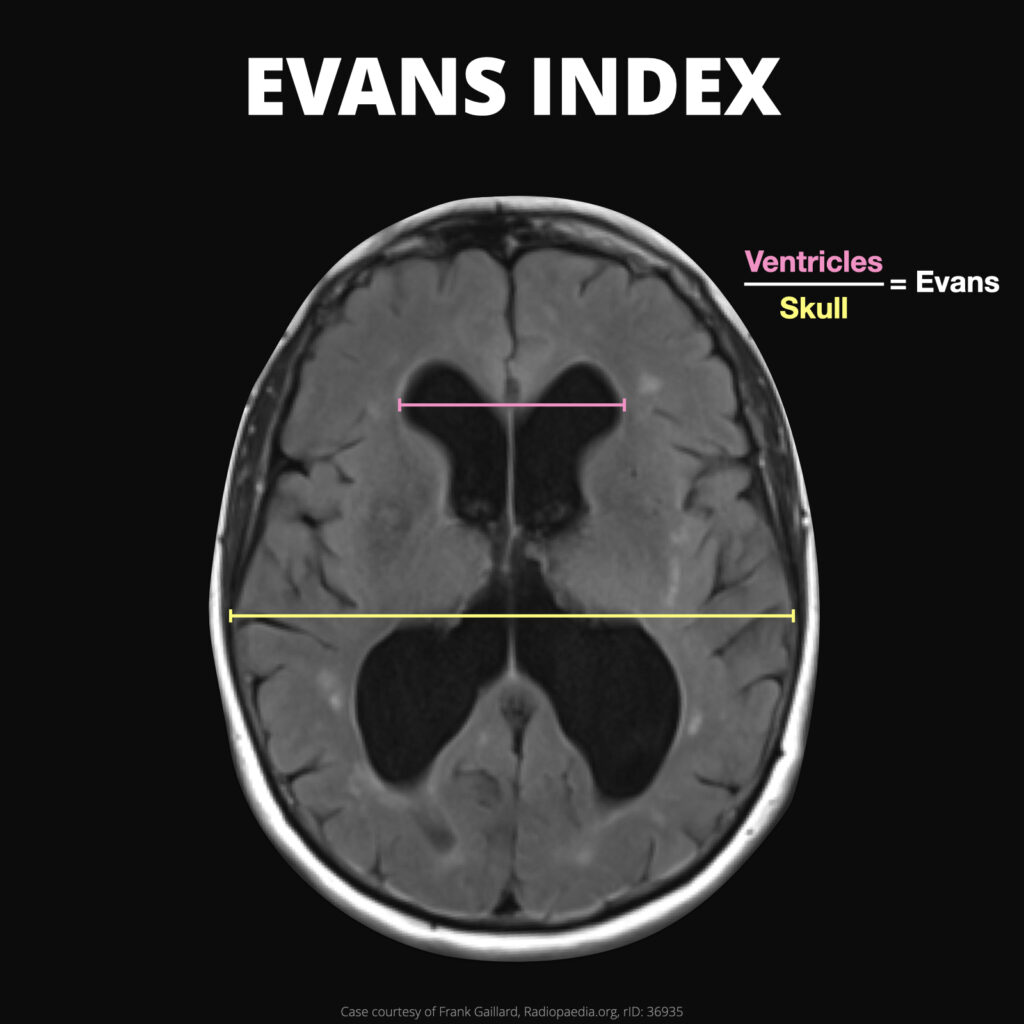
- Ventricular enlargement (Evans’ index >0.3 — Evans index is the ratio of the maximum width of the frontal horns of the lateral ventricles and the maximal internal diameter of the skull at the same level employed in axial CT and MRI images).
- Temporal horn dilatation = early sign.
- Ballooning of frontal horns.
- Transependymal CSF seepage → periventricular hypodensity.
- Underlying cause:
- Intraventricular hemorrhage.
- Mass lesion/obstructive tumor.
- Aqueductal stenosis.
Case courtesy of Frank Gaillard, Radiopaedia.org. From the case rID: 165931
🧲 MRI in Hydrocephalus
Advantages
- Superior soft tissue and CSF flow imaging.
- Better for etiology and classification.
- No radiation (important in pediatrics).
MRI Findings
- Same as CT for ventricular dilatation but with added value:
- Sagittal T2 → excellent for aqueduct and 4th ventricle outflow.
- Cine phase-contrast MRI → CSF flow dynamics (obstructive vs communicating).
- Periventricular hyperintensity on T2/FLAIR = transependymal seepage.
- Detects arachnoid cysts, Chiari malformations, Dandy-Walker, posterior fossa tumors.
📊 CT vs MRI in Hydrocephalus (Comparison Table)
| Feature | CT | MRI |
|---|---|---|
| Speed | Fast, emergency use | Slower, not ideal in unstable patients |
| Radiation | Yes | None |
| Ventricle size | Excellent | Excellent |
| Cause detection | Limited (mass, bleed) | Superior (tumors, congenital malformations, webs) |
| CSF flow assessment | Not possible | Cine phase-contrast MRI useful |
| Pediatric use | Limited (radiation risk) | Preferred |
| Best use case | Acute hydrocephalus, trauma, emergency bleed | Etiology workup, surgical planning, chronic hydrocephalus |
📝 Teaching Points
- CT → best for acute emergency diagnosis.
- MRI → best for etiology, CSF flow, congenital anomalies, surgical planning.
- Always look for cause of obstruction, not just ventricle size.
✅ Conclusion
CT is the frontline modality for rapid detection of hydrocephalus and its acute causes, while MRI is the gold standard for identifying the etiology and guiding management. Together, they complement each other in the diagnostic pathway.